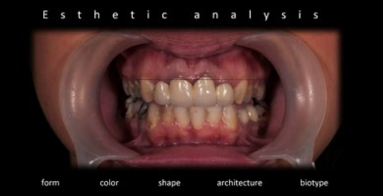
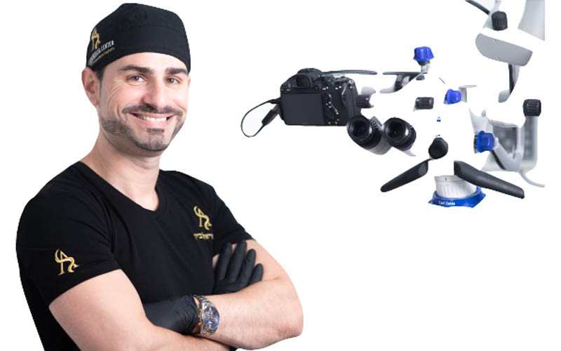
Microscope as precision tool – Clinical complex case report

Share This Page

Clinical case presented by Dr. Ariel Savion, D.M.D, LL.B, M.Sc, I.C.O.I

Case History

A complex case of rehabilitation in the upper jaw with old porcelain-fused metal crowns in teeth 1.2 – 2.2. This case concerns a 29-year-old patient who complained about unaesthetic and poor dental brilliancy. During the clinical interview, the patient stated that evidence of this can be found in the intra-oral and dento-labial image as well as in the patient’s facial portrait. All images were required to generate a correctly analyzed aesthetic preview. The  hole case was treated under the ZEISS EXTARO 300 microscope (ZEISS, Germany) to maximize precision in aesthetic outcome.

Magnification avoids unnecessary destruction of healthy dental tissue. With a microscope, it is possible to see boundaries between restorative material and dental tissue in great detail. 

Fig. 1 - Initial situation during facial analysis, including teeth exposure in smile and functional video.

Case report

Clarity leads to conservative tooth preparation and avoids loss of healthy tissue and pulp damage. Aesthetic outcomes depend on the harmonious integration of fundamental aesthetic criteria with the smile and character of the individual. Ideally, the outline of the gingival margin should be parallel to the incisal line and horizontal to the reference line. Photographic documentation is important for correct aesthetic assessment, especially in complex cases such as this one, giving the patient strong motivation to undertake the treatment.

Fig 2. - High smile line with uneven smile. High gingival exposure.
Fig 3. - Dento-labial analysis with extra oral examination.
Fig. 4 - Gingival analysis in profile smile.

The patient presented inflamed tissue with reddish color. Teeth 1.2, 1.1, and 2.1 were treated endodontically. It was observed that there was thin periodontal tissue with exposure of clinical crowns. In clinical examination, bleeding was observed, proving poor oral hygiene.

The appearance of gingival tissue (Fig. 6,7) plays an important role in the overall aesthetic framework, especially in patients with an average or high smile line. The goal is to restore an ideal outline of the gingival margins under magnification for better precision. Nevertheless, adequate biological integration of restorations has to be maintained.

Page 1
Fig. 5 - Gingival analysis in profile smile. One can observe gingival recession and high smile line. The appearance of gingival tissue plays an important role in the overall aesthetic framework.

The use of magnification allows maximum restoration longevity, such as marginal integrity, avoiding gaps, and excessive material. Magnification helps reduce failures, such as secondary caries, fracture, marginal deficiencies, wear, and postoperative sensitivity.

 For durability of restorations, we need to follow the biomechanics aspects,  redictability, magnification, and illumination. New materials and techniques have developed in recent years and continue to improve on a daily basis.  

Restorative procedure becomes more complex and sophisticated and requires  ore focus and attention. Hence, the use of the microscope nowadays in minimal invasive dentistry is mandatory for improving our patients’ lives for the better. Moreover,  agnification will change the professional dentist’s life with more predictability and precision in indirect restorations.

Fig. 6 - Aesthetic analysis with gingival recession in teeth 1.1 and 2.1 with poor marginal integration.
Fig. 7 - Gingival margin outline with disharmony in gingival margin. High gingival exposure.

The appearance of gingival tissue (Fig. 6,7) plays an important role in the overall aesthetic framework, especially in patients with an average or high smile line. The goal is to restore an ideal outline of the gingival margins under magnification for better precision. Nevertheless, adequate biological integration of restorations has to be maintained.

Fig. 8 - Gingival margin asymmetric. Thin gingival biotype with gingival inflammation.

Microscopic Examination – In the initial situation prior to starting the treatment plan, the magnification under the microscope is particularly useful for examination of the
external surface of the tooth. Caries can be identified in areas that are difficult to see without magnification under Fluorescence Mode (ZEISS EXTARO 300, ZEISS, Germany).
Fluorescence Mode is intended to be used by qualified physicians in dentistry as an aid in the detection of dental caries and as an aid to distinguish composite resin material
from natural dentin enamel. 

Tooth Analysis – In the presented case, gingival margin was asymmetric with irregular zeniths of gingiva. Nevertheless, thin biotype, absent papillae with gingival inflammation was observed. Tooth proportion, contour, arrangement, axes are abnormal. 

Bacterial Plaque Control – An imperfect surface provides favorable sites for residual plaque deposition. The process promotes caries development and periodontal diseases. Rough or over-contoured surface can shorten the longevity of direct to indirect restorations.

Page 2
Fig. 9 - Pre-operative x-ray prior to starting treatment plan.
Fig. 10 - Microscopic examination with increased pathogenicity of subgingival plaque, inadequate margin and contours with presence of discrepancy.

Once all the photographic and clinical evidence has been collected, the dentist starts constructing the wax-up analysis. Wax-up analysis was carried out with reference to the study cast. A silicon template was made according to the wax-up model for gingival correction (Fig. 11,12).

Fig. 11 - Diagnostic wax-up as step of initial information prior to starting treatment plan.
Fig. 12 - Silicon template was produced according to wax-up model, for gingival correction under magnification.

In situations of a high smile line, old restorations did not extend the subgingival level. Therefore, there was no need for respective surgery. Crown lengthening surgery includes added expense, patient inconvenience, and discomfort. Moreover, it may compromise aesthetics and support of adjacent teeth. Crown lengthening surgery is utilized to create more available vertical tooth structure for crown retention, especially in situations of biological width violation and integrity.

Fig. 13 - Gingival correction using silicon template for adequate symmetric gingival margin.
Fig. 14 - Teeth 1.1, 2.1 with insufficient gingival margin and violation of biological integration. In case of high smile line, no need for soft tissue augmentation. Gingivectomy sufficient for gingival correction.
Page 3

One should realize that violation of biological integrity occurs due to inadequate marginal closure, incorrect coronal contour, surface roughness, and access of cement. Consequently, magnification is really useful to avoid unnecessary destruction of healthy dental tissue. Under the microscope (EXTARO 300, ZEISS, Germany), it is possible to see the boundaries between the restorative material and dental tissue in great detail. Without magnification, this cannot be seen with the same clarity, leading to more extensive tooth preparation, loss of healthy tissue, and potential damage of the pulp in vital teeth.

Fig. 15 - Old PFM crowns removed under magnification in the same appointment afterwards.
Fig. 16 - Marginal configuration inevitably affects esthetic result and degree of resistance of definitive restoration.

Restoring teeth functionally and aesthetically after endodontic treatment often presents a challenge to the dentist. In the case presented, after the old PFM crowns are removed, one can observe extensive loss of tooth structure that reduces structural integrity. Substantial plaque and caries lesions with gingival inflammation due to inadequate marginal adaptation as well as accelerated caries lesions and increased phatogenicity of subgingival plaque. The aim of the clinician and technician is to achieve excellent margins and perfect adaptation. Clear visibility of the surgical field and high magnification are very important to reach that goal.

If excellence is achieved, the results are highly satisfying for both the patient and the clinician, and also for long-lasting indirect restoration.

Fig. 17 - Violation of biological integrity with insufficient mechanical retention of prosthetic.
Fig. 18 - Replacement under magnification, avoiding impairment.

When we replace restorations (aesthetic or non-aesthetic) due to a recurrent caries lesion, healthy tooth material is often also removed at the same time. Recognizing the limits between teeth and restorations, seeing these structures with magnification and high-quality light, means greater preservation of tooth tissue. Reduction of structural integrity of the teeth leads to a decreased resistance to fracture. Improving lighting coupled with magnification provides a clear distinction between surface, decay, dentine, enamel, composite, posts, and marginal adaption.

Fig. 19 - Teeth structure with inadequate resistance and retention form under magnification after caries removal.
Page 4

While endodontically treated teeth may fail for a variety of reasons, such as caries, endodontic failure, and periodontal disease, among others, tooth fracture remains a significant factor in the post-treatment extraction of these teeth. Endodontically treated teeth can be restored with directly placed restoration in the anterior region.

Fig. 20 - Severe damaged tooth loss, patient only 29 y.o. Without magnification, saving the teeth from extraction, was impossible.
Fig. 21 - Tooth structure remaining. Occlusal view under magnification.

Before restoration with a massive loss of tooth structure due to invasive and unpredictable treatment, some factors have to be considered, such as systemic health, periodontal condition, caries susceptibility, tooth function, tooth structure remaining, materials factors, type of definitive material, post type, core, cement.

Fig. 22 - Risk analysis under magnification before direct abutment restoration.

The risk analysis should be taken into account before abutment of direct or indirect restoration. Presence of radiographically dense and adequately extended obturation must be confirmed before continuing to the next step. Furthermore, absence of symptoms and periapical pathology and the quality of existing endodontic treatment have to be assessed, and periodontal disease controlled. In the presented case, the root canal has not been exposed to oral environment, and the crown-root ratio has been assessed.

Fig. 23 - Tooth 2.2 was treated endodontically due to root canal infection.
Fig. 24 - Silicon key displays massive loss of tooth structure before restoration.

Rubber dam recommended, especially under magnification. Although, patient with respiratory deficiency refuses to isolation under rubber dam. Therefore, in the presented case, a retraction cord was used for better control in direct restorations. A silicon key (Fig. 23) based on the diagnostic wax-up demonstrates massive loss of tooth structure.

Fig. 25 - Material chosen - Post quarts fiber - DT light post, BISCO, JAPAN.
Page 5
Fig. 26 - Resin composite - Gradia Core, GC, JAPAN.

Material Selection (Fig. 26) – Tooth restoration was performed with quarts fiber post (Bisco, Japan). “DT Light” post shows elastic modeled (GPa ) 42, flexural strength (MPa) 1,131, lower incidence of root fracture. It provides the best combination of strength and favorable modulus. The cementation can be performed with a self-curing mode that reduces stress and avoids tooth fracture. In choosing resin composite, Gradia Core (GC, Japan) shows excellent mechanical properties: bond to tooth structure, dual-cure and easy-to-use, shows great resistance to fatigue loading, favorable in aesthetic consideration as tooth color material, and has fracture toughness. Ferrule effect must be respected to reinforce the remaining tooth structure and help the restoration withstand lateral forces.

Fig. 27 - Key factor - adequate ferrule for definitive restoration and fracture resistance.
Fig. 28 - Ferrule provides resistance form and enhances longevity.

Ferrule refers to the dimension of remaining vertical tooth structure from the anticipated margin of the preparation to its coronal extent, which is available for encirclement by the crown. Furthermore, the ferrule effect serves to reinforce the remaining tooth, provides resistance form, and enhances longevity. The circumferential ferrule must be at least 2 mm, otherwise the chance of failure increases dramatically.

Fig. 29 - Tooth preparation under magnification, based on diagnostic wax-up.

At any rate, the biological width must be respected. The definition of the biological width-dimension of junctional epithelium and connective tissue attachment to the root above the alveolar crest has been estimated to be approximately 2 mm.

Fig. 30 - Respect biological width before definitive restoration

A lack of adequate ferrule increases the risk of failure because of the post-crown dislodgment post-fracture and root fracture. A ferrule of only 1 mm in vertical height (Sorensen and Engelman) doubles the resistance to fracture of a tooth without remaining coronal tooth structure. Dental preparations were carried out with slight chamfer margin using calibrated finishing drill to obtain a reduction of 1.5 mm on the incisors (Fig. 29).

Page 6
Fig. 31 - Biological width estimated to be approximately 2 mm.
Fig. 31 - Final teeth preparation before final cementation. Precision is the key for restoration longevity.
Fig. 32 - Teeth 1.3, 2.3, 25, 26 - without endodontic treatment. Preparation wase performed under magnification of microscope EXTARO 300, ZEISS, Germany. Occlusal view.

The microscopic approach improves tissue preservation and handling while using specific techniques. Modern dentistry is based on precision, and precision is an absolute must to achieve high quality standards. Some form of magnification is indicated in all fields of dentistry, and not only in endodontic treatments. Magnification should be used by all dentists, in all areas. Dentists today demand the intensive utilization of computers and many other new technologies to aid the management of all the clinical information, digital documentation and record-keeping generated during the diagnosis and execution of clinical cases. Dentistry today demands a multidisciplinary approach in contrast
to the unilateral perspective of the past. Nowadays, microscopic dentistry is an “innovative” way to see and do dentistry. The treatment philosophy is now changing to a

Fig. 33 - Final teeth preparation.

more conservative approach, and the concept of minimal intervention is gaining popularity in modern dentistry throughout the world. In a long term, direct restorations as presented in this case allow the maximum longevity of the restorations, and marginal integration is the first point to be analyzed for measuring this success.

Fig. 34 - Definitive indirect restoration should correspond to esthetic parameters.

Biscuit Try In – Aesthetic parameters must be respected. Definitive restoration should correspond in volume and tooth position. Provisionals represent the prototype for definitive prosthesis in all aspects. Definitive restorations achieved from provisional restoration in function.

Page 7
Fig. 35 - Facial and dentolabial aesthetic parameters.
Fig. 36 - Before and after whilst lip in rest.

In these images (Fig. 35, 36), the final result of the prosthetic restoration can be assessed. Biological integration is highlighted together with satisfactory functional and aesthetic integration of the rehabilitation.

Fig. 37 - Integration of function, biological, aesthetic parameters should be respected.
Fig. 38 - Photographic documentation in profile during smile, before treatment, and final.

Photographic documentation in final result corresponds to aesthetic parameters and natural outcome. Horizontal plane corresponds to commissural and intra-pupillary lines (Fig. 35, 36). Documentation is essential in dentist practice, especially when dealing with aesthetically complex cases. DSLR camera or full frame is recommended. Furthermore, microscopic documentation under the microscope, leading dentist to document diagnosis and treatment, whether for forensic purpose, for the dentist’s own documentation needs, patients’ educations, training or case presentation.

Fig. 39 - Gingival level maintains adequate parallelism with occlusal plane and horizontal reference lines.
Fig. 40 - Smile line with ideal tooth position and arrangement.

Parallelism – Outline of gingival margin (delineated central and canine) should be parallel to both the incised edge and the curvature of lower lip.

Symmetry – Gingival margin of centrals and canine should be symmetrical and more apical position compared to laterals.

Page 8
Fig. 41 - Final outcome – follow-up after 3 months with symmetry, parallelism, papillae.
Fig. 42 - Final outcome - before and final outcome. Saving teeth from extraction.
Fig. 43 - Post op x-ray. One can observe the perfect adaptation and precision.

Summary

In the presented complex case, many of the procedures highlighted could not be performed without magnification. Therefore, it enables the clinician to offer many more treatments than otherwise possible. Under magnification, clinicians have more control of the clinical environment, which can lead to greater efficiency, less stress and more predictable outcomes. Patients immediately come to understand that dentists using a microscope work in high- level treatment standards. The documentation of the whole procedure also increases acceptance of treatment.

Clinical photography is essential for communication between the clinician and technician. Furthermore, documentation is essential for patient documentation, especially in the aesthetic field. Microscopic documentation provides a powerful communication tool. Whether it is still or video images, showing patients the state of their oral health makes it easier for them to understand the problem at hand.

Page 9

Health benefits of the dentist

Improved ergonomics and posture with the microscope EXTARO 300 by ZEISS become more obvious as the dentist becomes more proficient in its use. Improved ergonomics reduce back and neck problems. Dentists have better ability to provide more treatments throughout the day/week. Better health results in a better work and better passion in the dental field.

Working under magnification provides many possibilities for viewing the surgical field. In restorative and prosthodontic dentistry treating in high-level standards achieves better durability in the conservative approach. Magnification
can improve our patients’ lives for better and change the dentist’s professional life. See more, do better. Precision is the key of success in the presented case. The 29 y.o. patient can smile without concern of uneven smile or any pain.

Page 10

Complete the form and a representative will contact you shortly

השאירו פרטים ונחזור אליכם בהקדם​