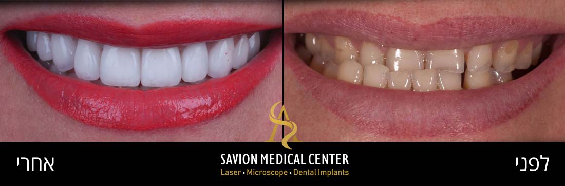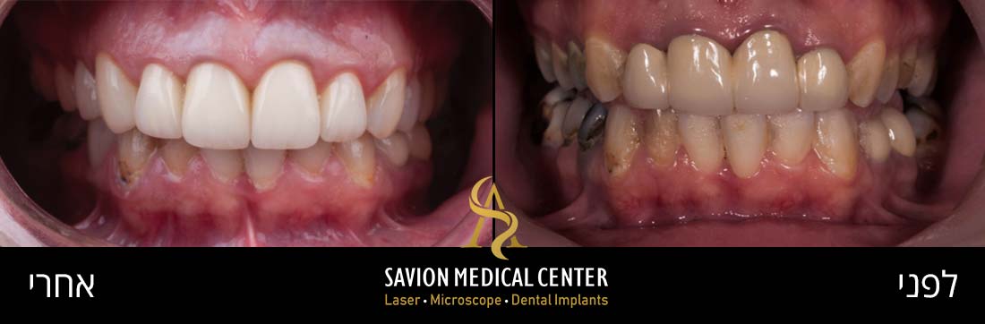
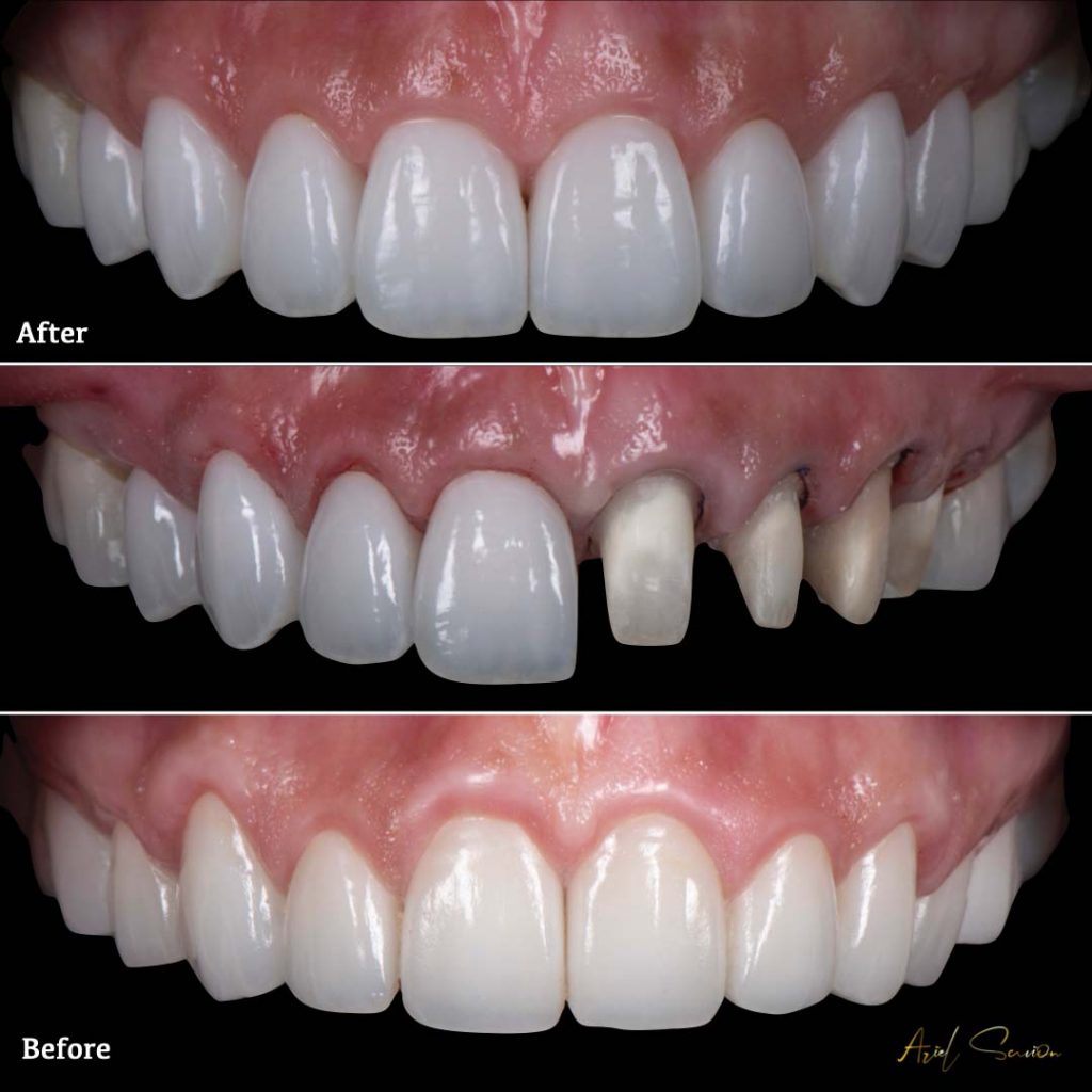
What Are Porcelain Veneers?
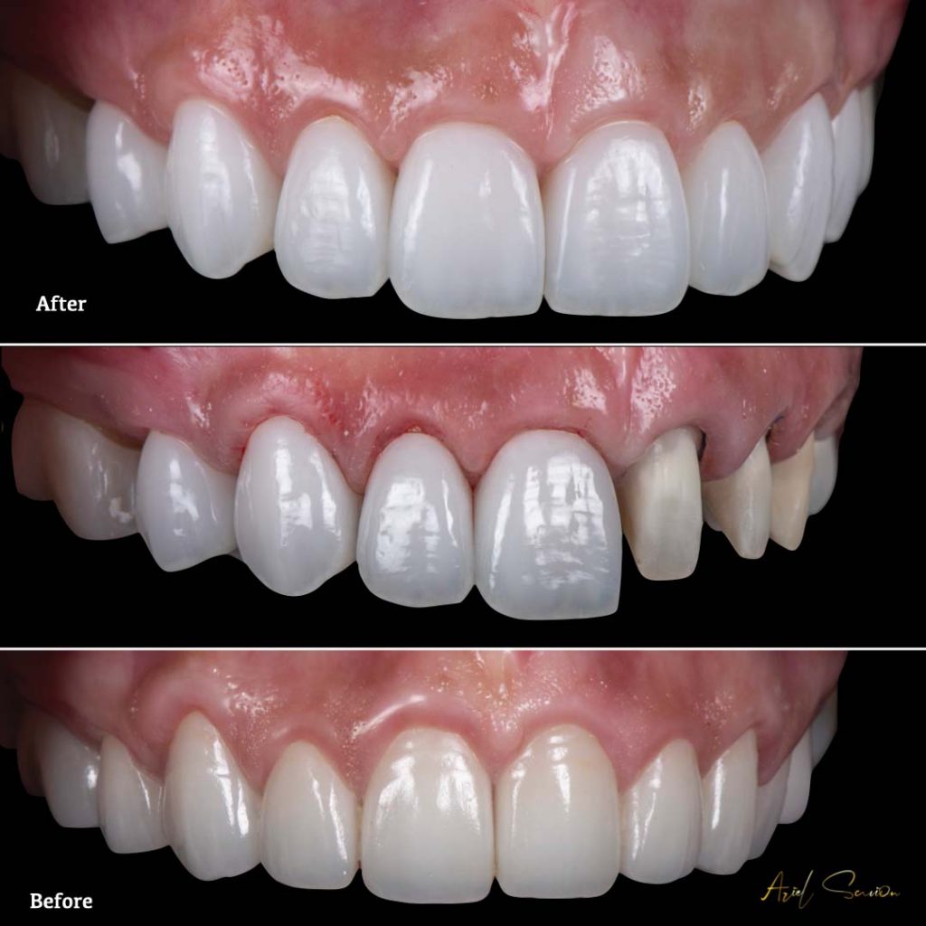
Porcelain veneers are a minimally invasive cosmetic dental solution in which a thin ceramic shell is bonded to the front surface of a tooth to improve its appearance or reinforce its structure. This treatment is commonly used to correct aesthetic imperfections and restore a natural-looking smile. Porcelain veneers are favored for their durability, translucency, and ability to closely mimic the appearance of natural teeth.
Veneers are typically made from feldspathic or monolithic porcelain and range in thickness from 0.2 mm to 0.7 mm. These custom-crafted restorations are bonded to the enamel, offering both cosmetic enhancement and functional protection.
There are two main types of veneer restorations:
- Indirect porcelain veneers, fabricated in a dental laboratory and bonded to the teeth in a subsequent appointment. These often involve a layering technique or staining to match natural dentition.
- Direct composite veneers applied chairside by the dentist using tooth-colored resin. These are less durable and aesthetic than porcelain alternatives but suitable for certain cases.
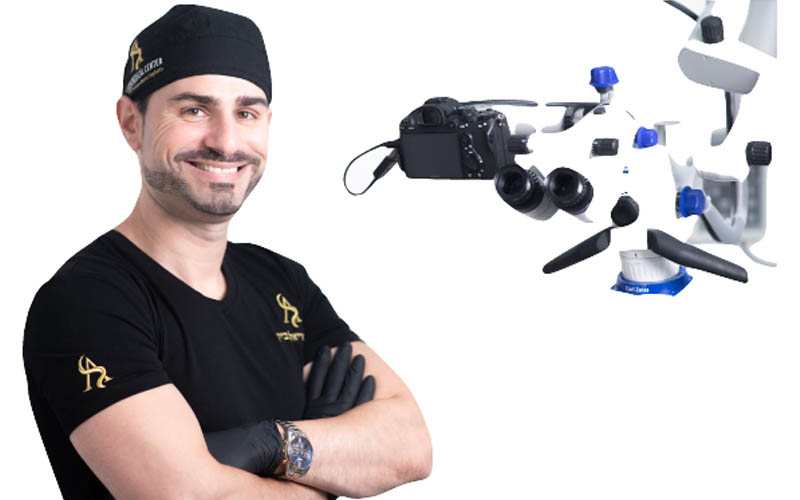
Dr. Ariel Savyon
Dentist since 2007, holds a double master's degree (Germany) in laser science and implantology (dental implants).
Serves as medical director of the dental corporation "Savyon Medical Center Ltd. Owner of a prestigious study club master_implant, treating dentists in various fields. The only certified instructor in Israel of the World Clinical Laser Institute in the field of laser science in dentistry in Israel.
Dr. Savyon, an opinion leader for leading companies in Israel and around the world, a researcher and international lecturer in the field of laser dentistry, periodontal medicine, and dental implants.
Who Is a Candidate for Porcelain Veneers?
Porcelain veneers are ideal for patients seeking aesthetic improvement to their smile. Veneers provide a harmonious, balanced smile that integrates naturally with the lips, gums, and facial features.
The treatment is elective and typically involves minimal reduction of enamel—ranging from 0.3 mm to 1 mm—though in certain cases no reduction is needed. Bonding to the enamel ensures superior adhesion and reduces the likelihood of veneer failure.
This procedure is commonly indicated for:
- Discolored or stained teeth unresponsive to whitening
- Teeth with irregular shapes or proportions
- Minor tooth misalignment, asymmetry, or gaps
- Chipped or worn teeth
Patients express appreciation for Dr. Ariel Savyon and the clinic team:
Aesthetic Benefits of Porcelain Veneers
Porcelain veneers offer a reliable cosmetic enhancement for patients aiming to improve tooth color, shape, and alignment without extensive intervention. Their optical properties allow for a natural appearance that closely resembles enamel.
Veneers help patients achieve a more uniform and visually balanced smile. Esthetic advantages include:
- Color correction for intrinsic stains or discoloration that cannot be resolved through bleaching
- Reshaping of teeth to improve proportions, symmetry, and alignment in the smile arc
- Closure of diastemas (spaces between teeth) and masking of chipped or slightly rotated teeth
- Customization of veneer shape and shade to match facial features and individual smile dynamics
- High stain resistance, as porcelain is non-porous and does not easily absorb pigments from food or beverages
Functional and Oral Health Benefits
Although primarily aesthetic, porcelain veneers offer functional advantages that contribute to long-term oral health:
- Enamel reinforcement: Veneers act as a protective barrier against external wear, particularly in patients with early signs of erosion or attrition
- Improved resistance to mechanical stress due to the strength of modern ceramic materials
- Occlusal correction in select cases, where veneers can help achieve better bite relationships and reduce strain on the temporomandibular joint (TMJ)
- Reduced risk of caries in the restored areas, thanks to the smooth, sealed surface that is less prone to plaque accumulation
The Treatment Process at Savion Medical Center
- Comprehensive medical and dental history review
- Full aesthetic and functional analysis, including occlusion, periodontal health, and tooth positioning
- High-resolution studio photography and intraoral digital scanning
- Shade analysis and smile line assessment
- Data from the first visit is transferred to a dental technician
- A digital smile design is created using CAD software
- A 3D-printed diagnostic model and silicone stents are produced for use in the preparation stage
- A digital and analog mock-up is evaluated intraorally
- Minimally invasive preparation is performed under a microscope
- Tooth reduction is guided by rigid silicone stents to preserve enamel
- Digital impressions are captured using an intraoral scanner
- A provisional restoration is placed while final veneers are being fabricated
The third and final clinical stage involves bonding the custom-made veneers to the prepared teeth. At Savion Medical Center, this procedure is performed under strict isolation and magnification to ensure optimal results.
- The patient first tries in the final veneers to confirm shade, fit, and aesthetics
- Isolation is achieved using a rubber dam to prevent moisture contamination
- Under a dental microscope, each veneer is bonded using resin cement following a multi-step adhesive protocol
- All bonding surfaces are kept dry and uncontaminated to ensure durable adhesion
Microscopic magnification allows for accurate placement and marginal adaptation, minimizing the risk of failure due to leakage, shade inconsistencies, or debonding. Proper bonding is critical, particularly for ultra-thin veneers, where the underlying tooth color may influence the final result.
Laser-Assisted Removal of Porcelain Veneers
Although porcelain veneers are intended as a long-term restoration, there are clinical scenarios in which removal is required—such as bonding failure, subgingival staining, or changes in treatment strategy. Traditional removal techniques rely on rotary instruments, which pose a risk of enamel damage.
tions of Conventional Methods
- Lack of contrast between veneer, cement, and enamel increases the chance of over-preparation
- Rotary grinding is invasive, time-consuming, and often leads to irreversible loss of tooth structure
- Removing multiple veneers this way increases treatment time and complexity
Modern erbium lasers allow for controlled, non-destructive debonding. Laser energy targets the water molecules within the resin cement layer, causing it to break down without affecting the underlying tooth or ceramic structure. This technique is particularly valuable when veneer preservation is essential, enabling re-bonding within the same session and avoiding the need for new restorations.
Is Laser Use Safe in Dentistry?
Laser technology is widely used in modern dentistry for both hard and soft tissue applications. When used by trained professionals, lasers are safe, precise, and effective.
Safety Considerations:
- Dental lasers target specific tissues based on wavelength and absorption characteristics
- Built-in sensors and safety mechanisms help regulate power output near sensitive areas
- Protective eyewear and strict clinical protocols are mandatory during use
Although minor risks exist—such as soft tissue irritation if misused—these are rare when the operator is experienced and the equipment is properly calibrated. At Savion Medical Center, laser-based treatments are integrated into daily practice for their precision, reduced invasiveness, and faster healing times.
Top 10 Essential Tips By Dr. Ariel Savion - Before Undergoing Porcelain Veneer Treatment
- Tip 1: Ensure Healthy and Well-Contoured Gums
Gingival health and contour are foundational to achieving a natural-looking smile. Irregular or inflamed gums can compromise even the most precise veneer work. For optimal results, the gingival margins must be symmetrical, stable, and free of inflammation.
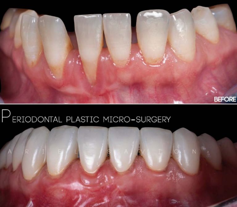
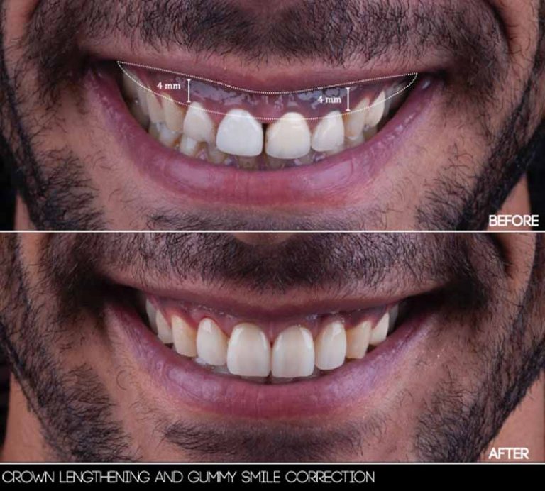
Gingival Plastic Surgery
Procedures such as crown lengthening, root coverage grafts, and frenectomy may be indicated to correct excessive gum display, recession, or soft tissue asymmetry. These interventions help create a balanced tooth-to-gum ratio that supports long-term aesthetic outcomes.
Treatment of Gingival Hyperpigmentation
Dark pigmentation of the gums, often due to melanin concentration or smoking, is common in patients with high smile lines. Although benign, it may be a cosmetic concern. Traditional treatments involving scalpels or abrasion are invasive and uncomfortable. At Savion Medical Center, laser ablation—using an erbium laser—is employed to gently remove pigmented epithelial layers, offering a safe and predictable solution with minimal discomfort and rapid healing.
- Tip 2: Do Not Skip Orthodontic Treatment for Misaligned or Protruding Teeth
Placing veneers on misaligned teeth without prior orthodontic correction often requires aggressive enamel reduction. This compromises bonding strength, as the adhesive performs best on intact enamel, which is highly mineralized. Over-preparation exposes dentin, a hydrophilic tissue that weakens adhesion and increases the risk of postoperative sensitivity and long-term failure.
Pre-Treatment Alignment
Orthodontic correction, even limited, often allows for conservative veneer preparation. For adult patients seeking a discreet option, Invisalign aligners can effectively align teeth prior to restoration. This approach supports both the biological and mechanical integrity of the final result.
- Tip 3: Insist on Professional Studio-Quality Photography
Clinical photography is not optional in modern aesthetic dentistry. High-resolution images taken under controlled lighting conditions are essential for diagnosis, treatment planning, patient communication, and quality control.
Applications of Professional Photography
- Accurate shade matching and documentation of baseline condition
- Evaluation of smile line, tooth proportions, gingival display, and facial symmetry
- Communication tool between the dentist and dental technician
- Reference point for before-and-after comparisons and patient education
At Savion Medical Center, procedures are also documented using 4K video via dental microscopes, enhancing diagnostic accuracy and treatment transparency.
- Tip 4: Request a 3D Digital Mock-Up Before Committing to Treatment
A diagnostic mock-up allows the patient and clinician to visualize the proposed smile design before any irreversible tooth preparation takes place. This step is essential for aligning expectations and identifying technical constraints in advance.
Digital Wax-Up and Analog Try-In
Using software, the ideal tooth shapes and dimensions are designed and printed as a 3D model. A silicone index is then fabricated and filled with provisional composite, which is applied to the patient’s existing teeth. After light curing, the patient can evaluate the aesthetics and function directly in the mouth. This mock-up is easily removed afterward without damaging the tooth surface.
- Tip 5: Choose a Clinic That Operates with a Full Digital Workflow
Digital dentistry enhances accuracy, efficiency, and communication throughout the veneer treatment process. Traditional impressions, wax-ups, and manual transfers are replaced with intraoral scanning, CAD design, and 3D printing.
Advantages of a Digital Workflow
- Faster turnaround time and reduced risk of human error
- Real-time data sharing with the dental lab
- Improved visualization of preparation margins and occlusal relationships
- Enhanced patient comfort with no physical impressions or trays
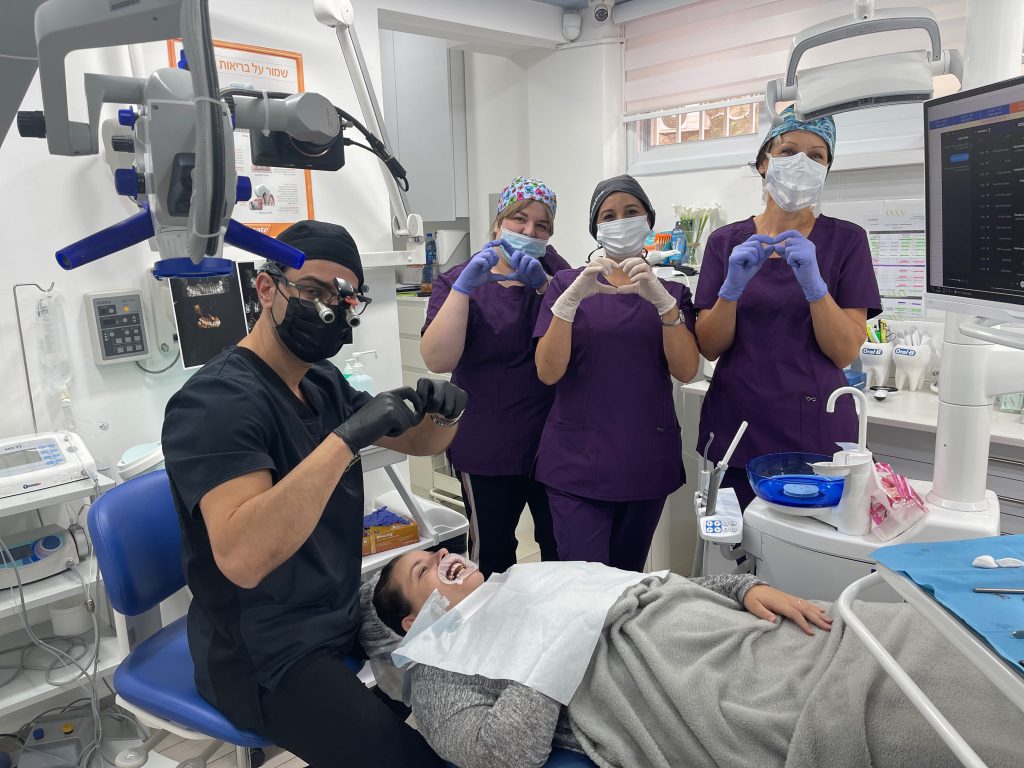
- Tip 6: Ensure Tooth Preparation Is Performed Under a Dental Microscope
Conservative preparation is critical for the long-term success of porcelain veneers. When reduction is performed without magnification, it increases the risk of unnecessary enamel loss, dentin exposure, and poorly defined margins. Enamel preservation is essential for achieving a strong, durable bond.
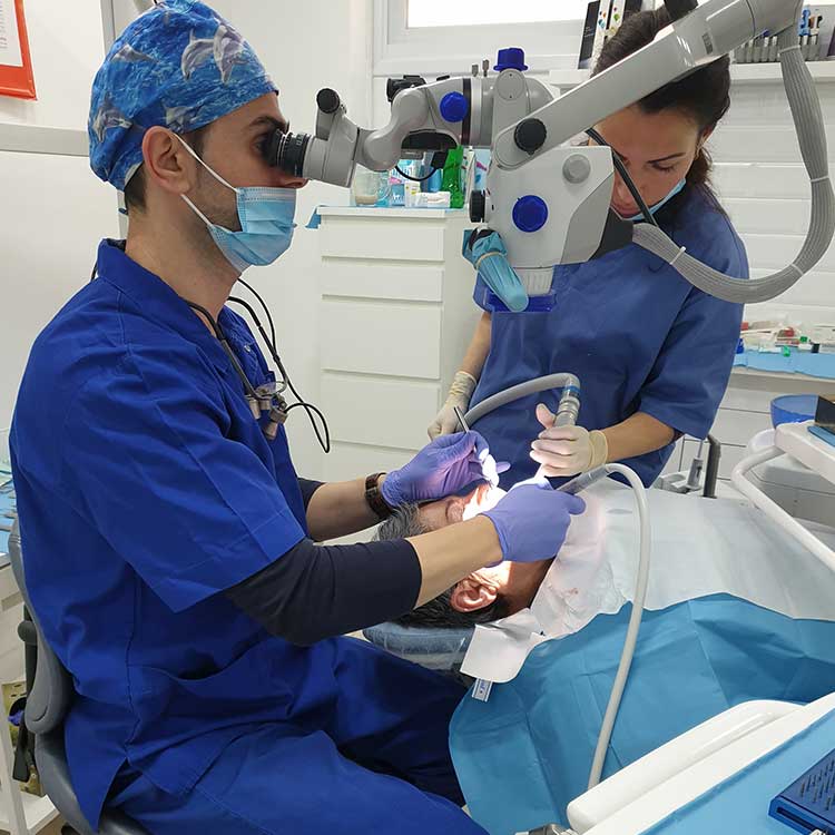
Microscope-Assisted Tooth Preparation
Using a dental microscope with up to 39× magnification, the clinician can precisely control the depth and extent of reduction, ensuring that the preparation remains entirely within enamel. At Savion Medical Center, tooth reduction is guided by 3D-printed stents derived from a digital wax-up. This method allows for minimally invasive, uniform, and controlled preparation.
Clinical Advantages
- Better visualization of margins and surface texture
- Increased bonding reliability due to enamel preservation
- Accurate finish lines that protect periodontal health
- Reduced need for future corrective work or veneer replacement
- Tip 7: Choose Laser Over Scalpels for Gingival Sculpting
In veneer cases, soft tissue contouring often plays a key role in achieving a symmetrical and natural-looking smile. Traditional scalpel techniques may cause bleeding, require sutures, and involve longer recovery times.
"Diode lasers used at Savion Medical Center allow for sculpting without anesthesia in many cases. Lasers also offer antimicrobial effects, enhancing soft tissue health during and after treatment."
Dr. Ariel Savion explains
Benefits of Laser Gingival Sculpting
- Precise tissue removal with minimal trauma
- Little to no bleeding, eliminating the need for sutures in most cases
- Faster healing and reduced postoperative discomfort
- Accurate definition of gingival margins, critical for impression-taking
- No risk of scarring or tissue shrinkage
- Tip 6: Ensure Tooth Preparation Is Performed Under a Dental Microscope
Conservative preparation is critical for the long-term success of porcelain veneers. When reduction is performed without magnification, it increases the risk of unnecessary enamel loss, dentin exposure, and poorly defined margins. Enamel preservation is essential for achieving a strong, durable bond.
Microscope-Assisted Tooth Preparation
Using a dental microscope with up to 39× magnification, the clinician can precisely control the depth and extent of reduction, ensuring that the preparation remains entirely within enamel. At Savion Medical Center, tooth reduction is guided by 3D-printed stents derived from a digital wax-up. This method allows for minimally invasive, uniform, and controlled preparation.
Clinical Advantages
- Better visualization of margins and surface texture
- Increased bonding reliability due to enamel preservation
- Accurate finish lines that protect periodontal health
- Reduced need for future corrective work or veneer replacement
- Tip 8: Veneer Bonding Must Be Performed Under Rubber Dam Isolation
Moisture control is a non-negotiable requirement during the bonding phase. Even trace amounts of saliva or blood can interfere with the adhesive process, leading to weak bonding, marginal leakage, and compromised aesthetics.
"At Savion Medical Center, bonding is performed under microscope magnification and rubber dam isolation to ensure that each veneer is seated precisely and sealed completely."
Dr. Ariel Savion explains
Why Rubber Dam Isolation Is Essential
- Provides a dry and clean field for bonding
- Prevents contamination that could alter the shade of thin, translucent veneers
- Improves the fit and long-term stability of the restoration
- Minimizes postoperative sensitivity by ensuring complete seal
- Tip 9: Verify the Skill and Artistic Ability of the Dental Technician
The final outcome of a veneer case depends not only on the clinician but also on the dental technician. Fabricating lifelike restorations requires a deep understanding of dental anatomy, light dynamics, and material science.
Qualities of a Competent Technician
- Expertise in ceramic layering and morphological design
- Proficiency in digital smile design tools
- Ability to match shades with natural tooth translucency and texture
- Close collaboration with the clinician throughout each case
"At Savion Medical Center, we works exclusively with master dental technicians to ensure that every veneer is fabricated to exact specifications. The dental technician’s attention influences the natural appearance, fit, and durability of the final result."
Dr. Ariel Savion explains

- Tip 10: Evaluate the Dentist’s Expertise and Commitment to Results
Choosing the right clinician is essential for achieving a successful and predictable veneer outcome. Veneer treatment is not purely cosmetic—it requires advanced knowledge in multiple dental disciplines, including occlusion, soft tissue management, material science, and digital workflows.
Qualifications and Clinical Philosophy – An experienced dentist will present a clear treatment plan supported by case studies, high-quality before-and-after photographs, and digital simulations. Attention to clinical detail—such as margin placement, preparation design, and shade matching—is critical for aesthetic and functional success.
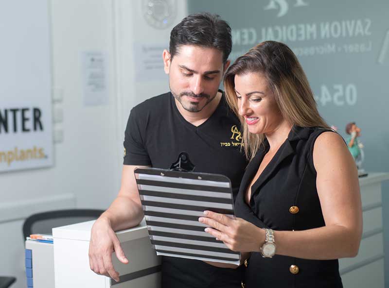
Clinic Environment as an Indicator of Standards – The appearance of the clinic and its staff reflects the level of care and attention given to each case. A well-organized, clean, and professional environment often signals a commitment to precision and patient satisfaction.
Transparency and Long-Term Responsibility – A trustworthy provider ensures open communication, realistic expectation setting, and long-term follow-up. Patients should feel confident that the dentist will address any complications or adjustments needed post-treatment without hesitation.
Long-Term Maintenance of Porcelain Veneers
Proper maintenance is essential for preserving the appearance and function of porcelain veneers over time. While veneers are durable, they still require regular care to prevent complications.
- Brush twice daily and floss regularly to reduce plaque accumulation
- Avoid biting on hard objects (e.g., ice, pens, or nutshells)
- Minimize intake of highly pigmented foods and beverages
- Wear a night guard if you grind your teeth or play contact sports
- Attend regular dental checkups for professional cleaning and monitoring
Why Dentist Selection Is Critical for Veneer Success
Each of the steps requires clinical experience and attention. Mistakes in any stage such as over-preparation or incorrect shade matching can compromise aesthetics, cause sensitivity, or lead to restoration failure. Success in veneer therapy depends on multiple technical factors:
- Tooth preparation must preserve as much enamel as possible to ensure optimal bonding strength
- Material selection must be tailored to the case, taking into account translucency, thickness, and underlying tooth color
- Bonding protocol must be carried out under ideal conditions using a microscope and rubber dam
- Shade selection requires coordination between dentist and technician, guided by digital photography and color-matching tools
Risks and Side Effects
While porcelain veneers are considered safe and effective, patients should be aware of potential risks:
- Tooth sensitivity, typically temporary, may occur after preparation or bonding
- Enamel damage can result from over-reduction
- Infection or inflammation is rare but possible if bonding is improperly performed
- Bite discrepancies may develop if veneers are placed without proper occlusal evaluation
- Material discoloration or edge wear can occur over time if inferior ceramics are used
What Defines an Attractive Smile?
Porcelain veneers enhance smile aesthetics in a natural, functional, and long-lasting way. An aesthetic smile is defined by balance among three primary components:
- Teeth – shape, alignment, proportion, and shade
- Lips – symmetry and framing of the dental arch
- Gingiva – contour, health, and exposure
Dental Photography and Data Transfer to the Laboratory
Professional Clinical Photography:
At Savion Medical Center, a dedicated in-clinic photo studio ensures consistent, standardized photography. This enables better planning and improves treatment outcomes.
High-resolution photography plays a central role in aesthetic dental treatment. It allows the clinician to document tooth color, shape, texture, gingival architecture, and lip dynamics with accuracy. These images serve several functions:
- Documentation for diagnosis and planning
- Visual communication during consultations – with the patient
- Precision in shade matching and design customization
- Alignment of expectations between the dentist and dental technician
Microscope-Enhanced 4K Video Recording
Using the integrated camera on the dental microscope, clinical procedures are recorded in 4K resolution. These videos document subtle intraoral details—such as shade nuances, margin definition, and soft tissue texture—and can be reviewed for quality control, legal documentation, or technician guidance in complex cases.
Still Photography Through the Microscope
Microscope-assisted still photography allows for high-magnification images of preparation surfaces, enamel texture, and bonding interfaces. This adds an extra layer of precision in documentation, patient communication, and interdisciplinary collaboration.
Advanced Digital Technology in Dental Treatment
CAD/CAM Workflow
Modern dental workflows rely on computer design and manufacturing to increase accuracy and efficiency. The process begins with an intraoral scan and continues through digital planning, model design, and fabrication of the final restoration using either milling or 3D printing.
Digital Workflow Benefits
- Elimination of traditional impression materials
- Real-time sharing of scans and planning data with technicians
- Enhanced visualization of preparation margins and occlusion
- Reduction in turnaround time and risk of human error
Diagnostic Wax-Up and Mock-Up Process
The process helps anticipate limitations—such as tooth width, length, and soft tissue visibility—and supports minimal intervention dentistry.
- Digital Wax-Up – A virtual model of the final smile is created based on clinical measurements and esthetic goals. It is then printed as a 3D model used for planning and reduction guides.
- Analog Mock-Up – The digital design is transferred into the patient’s mouth using a silicone index and temporary resin. This allows evaluation of aesthetics and phonetics in real-time before finalizing the treatment plan.
Digital vs. Conventional Impressions
Conventional (Analog) Impressions
Traditional impressions involve silicone-based materials and trays. These are sensitive to handling errors and material shrinkage, and may cause discomfort in patients with a strong gag reflex.
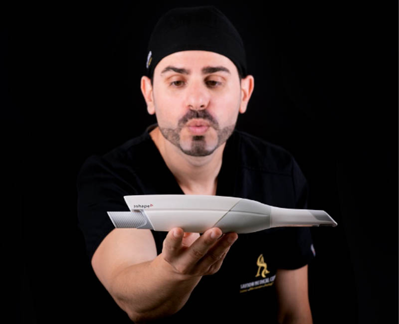
Digital Impressions
Using an intraoral scanner, a full-color 3D model of the oral cavity is captured and transmitted instantly to the lab. This method improves:
- Accuracy of details such as margins and interproximal spaces
- Patient comfort
- Workflow speed and reliability
- Consistency in results
Digital impressions eliminate the variables associated with analog materials and increase the overall predictability of the treatment.
Historical Perspective, Porcelain Veneers and Smile Design
The concept of smile aesthetics has evolved through centuries, rooted in the principles of proportion and symmetry. In classical antiquity, Greek philosophers introduced the idea of the “golden ratio”—a mathematical expression of harmony and beauty—which continues to influence modern design in various disciplines, including dentistry.
In 1978, Dr. Levin adapted these proportional guidelines to dental and facial structures. His work laid the foundation for modern smile design, where the ideal balance between tooth width, height, and spacing is calculated to achieve a visually pleasing result.