
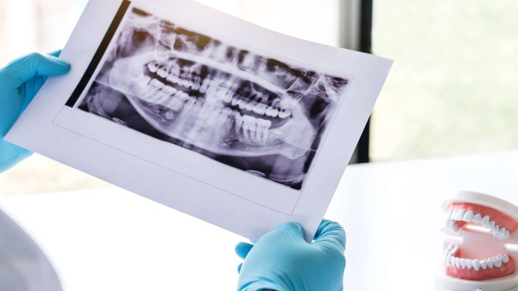
What is 3D X-ray Imaging - Dental CT?
Three-dimensional X-ray imaging, also known as CBCT scanning, is an advanced imaging technique currently used for diagnostic purposes in dentistry.
Unlike conventional X-rays (two-dimensional), which provide a "flat" image of dental structures and bones in general, 3D X-rays produce a three-dimensional result.
This technology uses a cone-shaped X-ray beam that rotates around the patient and generates multiple images from different angles. These images then allow for creating a three-dimensional representation of the patient's mouth, teeth, jaw, and additional areas of the face.
3D X-ray technology, sometimes referred to as "dental CT," undoubtedly represents a significant advancement in dental treatment. The resulting image provides the dentist with clear and detailed information about the entire oral cavity and teeth that would be impossible to discover through conventional X-rays.
The three-dimensional image reflects the precise shape and location of teeth, surrounding bone structure, connections between relevant tissues, and more, in a tangible and clear manner.
As we will elaborate further, these types of scans allow the dentist to plan and prepare for various types of treatments with an exceptionally high level of precision and preparation. All this is for the benefit of patients and the success of treatments.
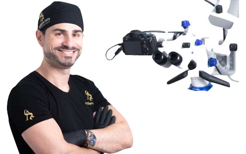
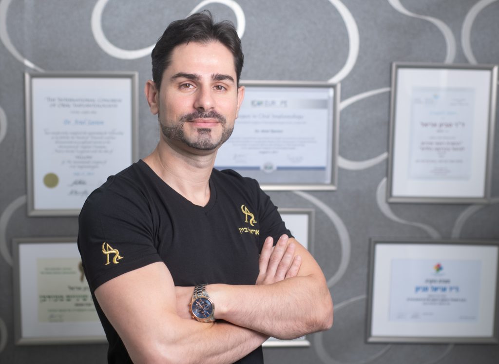
Dr. Ariel Savion has been an active dentist since 2007 and holds a dual Master’s degree from Germany in Laser Sciences and Dental Implantology. He has clinical expertise in laser periodontal therapy, microscopic surgery, and dental implant procedures.
He serves as the Medical Director of the dental corporation Savion Medical Center Ltd. and is the founder and owner of the prestigious master_implant educational club, dedicated to training dentists in advanced fields of dentistry.
In addition, Dr. Savion is the only certified instructor in Israel on behalf of the World Clinical Laser Institute in the field of laser dentistry.
Dr. Savion is an international researcher and lecturer and serves as a Key Opinion Leader (KOL) for leading medical companies in Israel and worldwide, specializing in laser dentistry, periodontology, and dental implantology.
The difference in patient experience between CBCT imaging at the clinic and regular CT scanning
Although there are certain similarities between CT and CBCT imaging, there is also a significant difference that affects the patient's experience during the scan. Many patients initially believe that CBCT imaging at the clinic will be performed lying down, as is customary with CT scans routinely performed in hospitals.
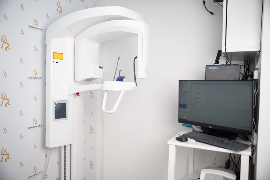
In fact, CBCT imaging, as performed in the clinic, is done with specialized imaging equipment with the patients standing (not lying down). Additionally, the entire process takes only about 17 seconds.
It's important to add that the scan involves less radiation compared to panoramic X-rays, and in some cases, the duration of radiation exposure can be reduced to less than 10 seconds.
| CBCT Scan | CT Scan | Parameter |
|---|---|---|
| Performed standing in the clinic | Usually performed lying down in a hospital | Scan position |
| Only about 17 seconds | Can last more than 17 seconds | Scan duration |
| Lower compared to panoramic X-rays; possibility of reduction to less than 10 seconds in some cases | Can be higher | Radiation amount |
Why is it important to have CBCT imaging in the clinic?
When CBCT imaging is available in a dental clinic, the level of care provided to patients can be significantly improved. The advanced technology of three-dimensional imaging allows for producing a detailed view of the mouth and assessing the patient's condition in the most comprehensive manner.
This type of imaging in the clinic allows the dentist to make correct and informed decisions regarding structural problems, dental and gum diseases, making it a valuable tool for therapeutic decision-making.
The three-dimensional images are produced in high resolution, allowing for detailed information about the anatomy of teeth, jaw, and adjacent dental structures to be inferred with unprecedented clarity.
At its best, the scan is so detailed that it can reveal bone structure, identify nerve pathways, distinguish some soft tissues, and even show interactions between tissues. The result is exceptionally high-level diagnosis, with the adaptation of a much more precise treatment plan.
CBCT scans allow for providing complex treatments based on reliable and accurate real-time information.
Based on the scans, dental implants can be placed, oral and maxillofacial surgical procedures can be performed, bone quality and quantity can be identified before implantation, complex root canal treatments can be performed efficiently and with great success, and more.
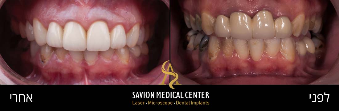
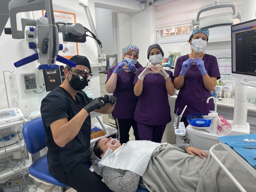
Patients express appreciation for Dr. Ariel Savyon and the clinic team:
Diagnosis and up-to-date CBCT imaging in the clinic
One of the most important diagnostic tools before beginning a dental implant process is computed tomography scanning (CBCT), thanks to its ability to provide detailed cross-sectional images of the implant site. With CBCT, accurate three-dimensional images of the oral and facial structures can be obtained.
CBCT scanning allows the dentist to discover critical details that might remain hidden in conventional X-ray imaging or clinical examinations. This non-invasive imaging method contributes to the understanding of bone quality, bone density, and relevant morphology, especially when assessing a patient's suitability for dental implant surgery.
As technology continues to evolve, CBCT scans are being updated and allow for enhancing the accuracy, predictability, and overall success of dental interventions. They have become an essential diagnostic tool for precise implant planning.
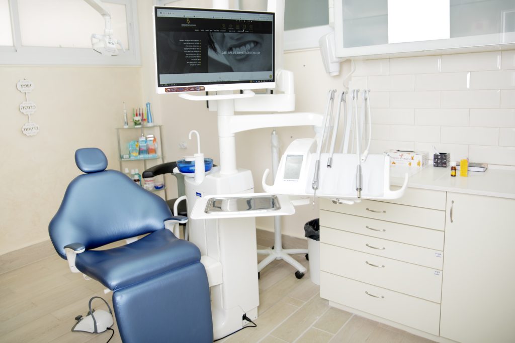
CBCT imaging at Savion Medical Center
At Savion Medical Center, we constantly strive to offer patients a wide range of services, all under one roof. As part of our service, you can come to the clinic for a clinical examination and have dental imaging done on-site using three-dimensional technology for some of the most advanced diagnostics in the world.
One of the significant advantages of performing CBCT imaging at the clinic is its accurate diagnosis, with which the dentist can propose to the patient a tailored treatment plan. The CBCT device at the clinic is a Japanese device from ASAHI and is considered among the most advanced in the world, with minimal radiation.
The CBCT scan radiation duration is a maximum of 17 seconds only. CBCT imaging is performed standing, not lying down, like panoramic X-rays with less radiation than panoramic X-rays. It is also possible to reduce the amount of radiation and radiation exposure to just 8 seconds.

First step
CBCT imaging
Second step
Data collection
Third step
Checking the results of the CBCT scan
9 Facts About CBCT Imaging
- adiation Standards in Israel - The Ministry of Health has established standards for the amount of radiation a patient may receive. General radiation in Israel is 2mSv, equivalent to 2000mSv. A CBCT scan of both jaws is equivalent to 0.2-0.3mSv. A person is allowed to receive 1mSv in one hour, provided they do not work in an area with radiation exposure.
- Flights Produce Radiation Similar to CBCT Scans!
- Types of Radiation - There are three types of radiation: alpha, beta, and gamma. Dentistry uses X-rays.
- Prescribed Dose in Israel - 0.1 mSv. 1.25mSv to 50mSv is considered dangerous; those who work with such risk levels are radiographers, as there is considerable radiation scatter in the room. This is why they have personal monitoring tags.
- Field of View - FOV - field of view. FOV is very important for imaging of the jaws, including the sinuses, in a single scan and obtaining as many details as possible. Imaging centers or dental clinics with CBCT machines should inform you about the type of machine including its FOV. A small field of view will hide many details such as nasal cavities, sinuses, etc., necessitating additional scans and exposing the patient to unnecessary radiation.
- Medium Field of View - The minimum required for 3D dental imaging in order to reduce the number of scans and radiation exposure.
- The Price of a CBCT Machine is determined, among other factors, by the range of the FOV, which is why not all CBCT machines function the same way. It's important to check the manufacturer, the range of the field of view, and the purchase date of the machine due to technological advancements.
- The Better the Resolution of a CBCT Machine, the Higher the Radiation Exposure.
- Currently, Advanced Machines Have Limitations on Radiation Amount - (Voxel - volumetric Pixel)
Guidelines During CBCT Imaging
CBCT scans allow for planning and performing complex, restorative, and preservative treatments with maximum precision. If you're scheduled to undergo dental implantation, periodontal disease treatment, or complex orthodontic treatment,
here are some recommended guidelines for the imaging session:
- Breathe Calmly
Make sure to breathe normally during the scan. There's no need for deeper than normal breaths.
- It's Advisable to Close Your Eyes
During the scan, it's advisable to close your eyes. Also, avoid swallowing saliva to prevent movement that could affect the quality of the scan. Remember that the scan is conducted in rotations and lasts less than 20 seconds.
- Dental Implants
For imaging and creating a surgical guide, soft tissues should be separated from the jaws by placing cotton rolls with the mouth not completely closed, preferably using a panoramic imaging device.
- For Orthodontic Treatment
The scan is performed with both jaws closed. Through this occlusion, many important details can be observed. During the exposure for imaging, the mouth should be closed without applying strong pressure on the teeth, as relaxation can cause movement.
How Does the Referral for CBCT Work?
According to the Ministry of Health's instructions, in order to provide a referral for CBCT scans, a comprehensive examination including the patient's medical history is required.
The guidelines state that the referring party must provide detailed justifications for why this type of scan is needed and why conventional X-rays are insufficient.
The referring physician must detail the reason for the referral, specify which areas they believe should be addressed in the scan, indicate the desired resolution, and so forth.

Interpretation of the Dental CT by the Dentist
Interpreting CBCT scans requires professional and thorough work by the interpreter or dentist. The interpretation process begins with selecting the appropriate imaging protocol for the case at hand. The protocol will correspond to the nature of the required treatment or the purpose of the examination.
After adjusting the field of view and resolution, the quality of the scan can be tailored to the area that needs to be covered, such as several adjacent teeth or a larger area in the face or jaw.
The interpretation is influenced by the quality of the scan, so it's important to ensure that patients follow the guidelines during imaging, such as patient waiting and no movement. When patients don't move during the scan, the interpretation will be easier and more accurate.
Additionally, the presence of metal fillings in the mouth may affect the quality of the scan and consequently the interpretation. Producing images for interpretation is related to the correct use of the machine, selecting the most appropriate types of images such as cross-sectional images, panoramic views, full 3D, etc. (all according to the nature of the clinical case at hand).
The interpretation will also take into account anatomical structures and findings from previous examinations and medical history, as relevant.
Types of Treatments that Utilize CBCT
Now we will elaborate more extensively on the types of treatments in which CBCT scans can be and are often very beneficial. These include, among others, treatments that require implants, periodontal treatments for patients with severe gum disease, oral surgery, and more.
In these types of treatments, CBCT imaging technology allows for a complete view of the anatomical structures in the areas where the implant is to be placed.
Such scans help identify bone quality and available bone quantity.
The imaging helps choose the optimal location for implantation and allows for extra caution to avoid damaging sensitive areas such as nerves or sinuses.
CBCT scans enable the dentist to make precise measurements and allow for surgical precision that contributes to safe and particularly successful implant surgeries.
Imaging of the bone structure in 3D technology can assist in selecting the type and size of the implant.
The scans also improve the dentist's preparation for implantation in a way that promotes success.
For patients requiring periodontal treatments, a CBCT scan can be very beneficial. Scanning with advanced technology provides detailed views of the level of damage or bone resorption, helping to assess the severity of periodontal disease.
Identifying the impact of periodontal disease on the bone helps plan the best treatment for the patients.
CBCT scans can produce good imaging of the gums, allowing for the identification of bone damage and additional damage, much more precisely than possible with regular X-rays.
The precision levels of the scans allow for targeted treatment for each case.
For example, treatments such as tartar removal, root planing, bone grafting, and so on. The result is improved quality of treatment and gum rehabilitation.
CBCT scans can also significantly assist in diagnosing infectious conditions in tooth roots, in preparation for root canal treatments.
The imaging allows for identification of the anatomical structure of the roots, detection of defects in the roots, as well as determining the extent of infection and its level of spread.
CBCT scans also allow for identifying the number of canals, their shapes, and any other relevant anatomical changes in the area to be treated. In this way, the dentist can offer particularly high-quality and precise treatment, even in complex cases.
In planning complex orthodontic treatments, the dentist may derive considerable benefit from CBCT scans. The three-dimensional imaging provides a panoramic view of the jaw, teeth, and bone structure.
The scan helps identify damaged teeth, diagnose the connection between teeth and jaws, and identify asymmetry that needs to be taken into account. The imaging is especially important in complex cases requiring very precise planning of orthodontic appliances, to create appropriate tooth movement throughout the treatment.
For oral and maxillofacial surgeries, CBCT scans assist in comprehensive diagnosis and subsequently in planning the surgical procedure. The detailed scans show the facial bones clearly, allowing the identification of defects, fractures, and so forth.
CBCT scans help in planning and performing jaw reconstruction surgeries, extraction of damaged teeth in complex cases, as well as treatment of temporomandibular joint disorders.
Benefits of Using Advanced 3D Imaging at Savion Medical Center
At your disposal at Savion Medical Center, advanced 3D imaging with a variety of benefits:
- Imaging for orthodontic treatment - Imaging before beginning the treatment for moving the teeth contributes to preventing bone resorption and gum recession.
- Imaging for preservation and prevention of extractions - Using imaging helps treat periodontal diseases and preserve teeth from extractions.
- In dental implants - Pre-operative or post-operative imaging helps prevent damage to anatomical structures.
- Better treatment of canals - Imaging in root canal treatments helps discover additional and hidden canals that might otherwise be "missed." Imaging also assists in better examination of infected canals.
- Imaging for better treatment of infections - The imaging allows for better diagnosis and treatment of various infection cases, such as in the sinuses, or temporomandibular joint infections.
- Imaging to improve complex extractions, for the benefit of patients - The imaging helps the dentist better prepare for complex extractions such as impacted wisdom teeth.
- To improve dental implant results - Imaging before bone implantation and ridge augmentation serves as a very important tool for installing dental implants with a perfect fit.
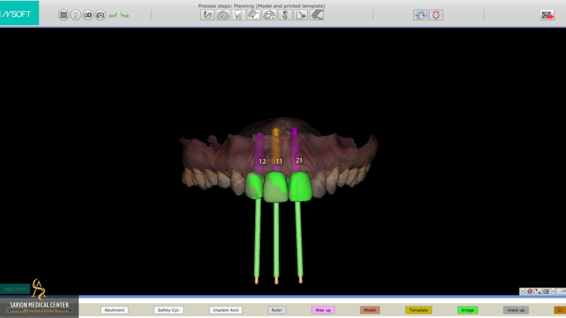
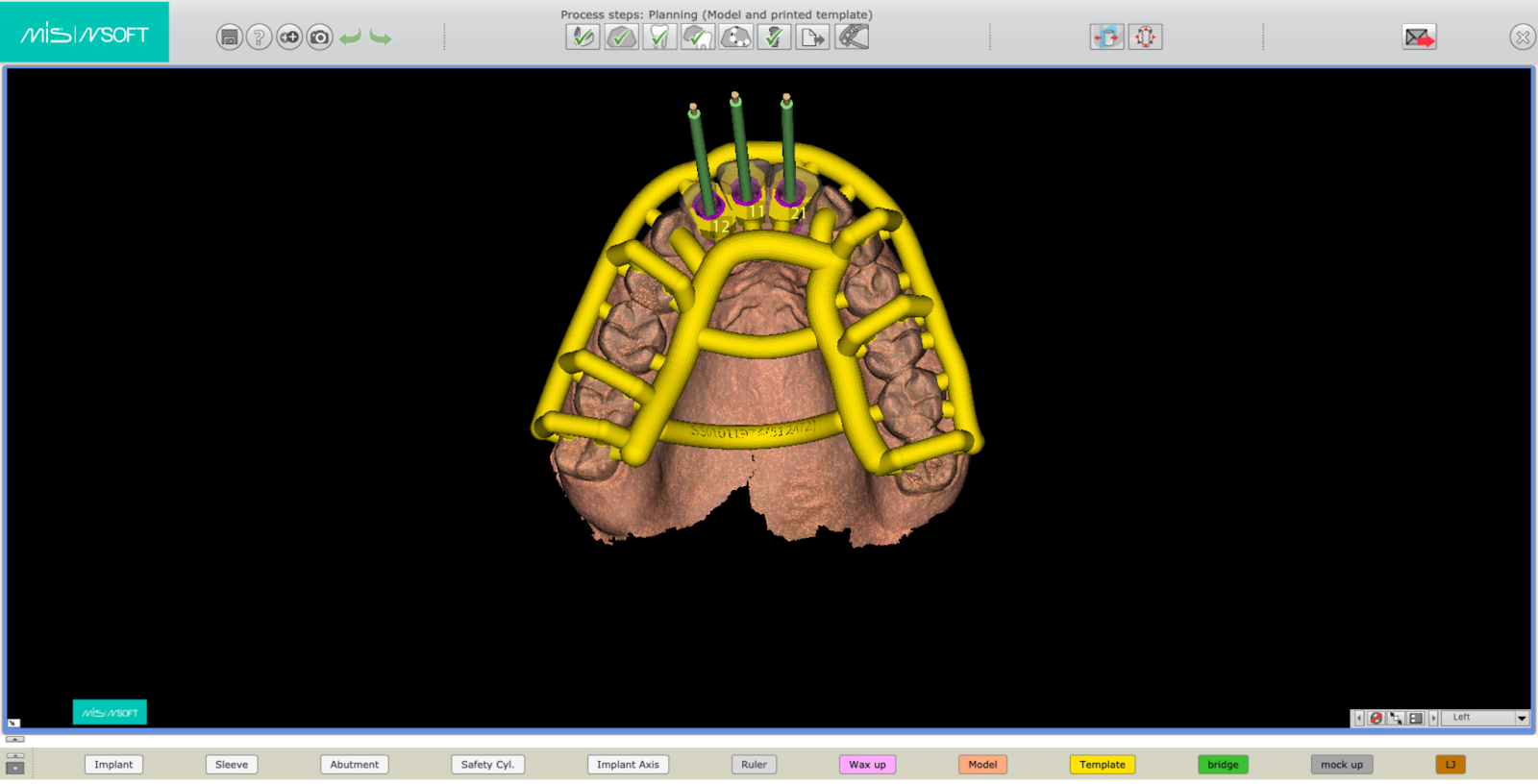
- Additionally, the imaging allows for the construction of a surgical guide as well as planning computerized surgery with unparalleled precision.

Microsurgery and Computerized Rehabilitation Using Advanced CBCT Imaging
- Microscope in Implantology – Using a microscope can significantly improve magnification, visibility, and lighting in the treated area. As a result, the implantation becomes more precise, the incision is minimal, and healing time is reduced.
- 3D Imaging – 3D imaging provides details of the jawbones, teeth, gums, and additional tissues. The imaging helps plan ideal locations for implantation and prevent peripheral damage.
- Computerized Surgery Planning with Breakthrough Software for Maximum Precision – Software tools combined with data obtained from CBCT streamline the implantation. One can use a simulation of the surgical results, plan where the implant will be placed, the position of the angle and depth, for the benefit of patients and the success of the implantation.
- Use of Light Energy – Laser – In dental implant surgeries as well as other treatments, laser plays an important role. This type of light energy allows making incisions in soft tissues with reduced discomfort and minimal bleeding, as well as disinfecting the area and improving healing. Lasers can be used to disinfect the implantation area, reduce the risk of infection, and more.
- Creation of a Surgical Guide through 3D Printing – 3D printing technology can assist in designing and manufacturing a surgical guide. Their purpose is to precisely fit the dental arches of patients during surgery, to guide the implant location accurately. This minimizes any chance of planning error and improves the outcome.
- 3D Printer – The printer also assists in preparing accurate dental models based on CBCT scans, for the production of crowns and additional dental solutions precisely tailored to patients.
- Intraoral Scan and File Creation – Scanning that presents in detail the structure of the patient's mouth and jaw. These digital models help design solutions for implants and dental rehabilitation. The process is considered very fast, highly accurate, and comfortable for patients, without the need for impressions using outdated, less comfortable, and less accurate methods.
Through CBCT imaging, innovative computerized surgery can be performed with advanced technology. The result is implantations and dental procedures that adhere well to the treatment plan, better patient experience, and high success rates in dental implants.